Must Reads
- What I learned Reading Hundreds of Strattera Reviews
- L-Tyrosine and Adderall: Read This Before Mixing Them
- Adderall Neurotoxicity: How To Protect Your Brain
- Focalin vs Adderall - A Side-By-Side Comparison
- Adderall Comedown? 10 Tips to Bounce Back After the Crash
- You'll Never Guess How Adderall Impacts Testosterone
 Oxidopamine (6-hydroxydopamine) – A toxic oxidation product of dopamine
Although Adderall confers clear clinical benefits in patients with ADHD, Adderall neurotoxicity is well-documented. Specifically, Adderall is neurotoxic to the dopaminergic system. Dopamine is a neurotransmitter that regulates reward, pleasure, motivation, and muscular tone.
Adderall improves attention by eliciting dopamine and norepinephrine release. Dopamine may auto-oxidize to form 6-hydroxydopamine which can damage dopaminergic neurons.
Dopaminergic neurons are particularly sensitive to oxidative stress18 due in part to their high metabolic rate. Parkinson’s disease is caused by the death of dopaminergic neurons in the substantia nigra. Methamphetamine use is considered a risk factor for the development of Parkinson’s disease.
Introduction: ADHD and Psychostimulants
The center for disease control (CDC) has estimated that over five million children have been diagnosed with attention deficit hyperactivity disorder (ADHD) in the United States alone. Psychostimulants, including amphetamine salts, are the most widely-prescribed medications for the treatment of ADHD.

State-based Prevalence Data of ADHD Diagnosis (2011-2012): Children EVER diagnosed with ADHD19
Despite considerable research in animal models and in vitro, the likelihood of irreversible Adderall neurotoxicity to dopaminergic neurons at therapeutic dosages in humans remains controversial and poorly characterized. Physicians have a responsibility to disclose the long-term risks of medications so that patients can make informed decisions with accurate cost-benefit analyses.
Adderall Neurotoxicity
The urgency of an accurate assessment of the risks Adderall neurotoxicity in humans is evident from the epidemiological finding that methamphetamine (METH) and amphetamine (AMP) abuse confers an increased risk of Parkinson’s disease 1. This ostensibly occurs due to Adderall neurotoxicity, i.e., the destruction of dopaminergic neurons or nerve terminals and global depletion of central dopamine.
Current research suggests that Adderall (mixed amphetamine salts) at therapeutic doses is highly unlikely to directly kill dopaminergic neurons. However, chronic amphetamine use does deplete endogenous antioxidants, results in long-lasting downregulation of the enzyme tyrosine hydroxylase involved in the endogenous biosynthesis of dopamine from tyrosine, and reduces both nerve growth factor (NGF) and brain-derived neurotropic factor (BDNF) in rat brain2. These cellular events likely underly Adderall neurotoxicity.
Benefits of Adderall in Patients with ADHD
The benefits of Adderall in certain patients cannot be dismissed despite these safety concerns.
An MRI-based neuroimaging study provided tentative evidence that therapeutic doses of psychostimulants in patients with ADHD rescues alterations in brain structure compared with unmedicated subjects3, which may underlie the clinical benefits of psychostimulants in this patient population.
It remains possible that amphetamine exerts neurotrophic effects in individuals with ADHD by facilitating normal brain development and enhancing reward saliency while also at the same time adversely affecting the dopaminergic system. Elevations in intracellular dopamine concentrations outside of vesicular storage likely promotes the formation of 6-hydroxydopamine (6-OHDA, oxidopamine). 6-OHDA or oxidopamine is a potent neurotoxin which selectively destroys dopaminergic neurons and is employed in animal models to emulate methamphetamine abuse and Parkinson’s disease.
These biochemical events likely underlie Adderall neurotoxicity.
Basic strategies to mitigate Adderall neurotoxicity
Vitamin C
Vitamin C is a blood brain barrier permeable antioxidant that seems to attenuate Adderall neurotoxicty. Vitamin C may help acidify the urine which promotes the clearance of Adderall. In addition, vitamin C is needed to synthesize dopamine. Therefore, in principle vitamin C supplementation could help regenerate dopamine synthesis and help ameliorate Adderall neurotoxicity.
The most cost-effective way to obtain Vitamin C is from PowderCity.
Vitamin D3
Supplement with vitamin D3. It has recently been reported that 1,25-dihydroxyvitamin D3 enhances endogenous glial cell-line derived neurotrophic factor (GDNF). In addition, exogenous treatment with GDNF reduces dopaminergic damage induced by 6-hydroxydopamine (6-OHDA) lesioning in animal models.
These two observations led Wang et al’s group to test the hypothesis that D3 might mitigate 6-OHDA induced neuronal injury. They found that 8 days of pretreatment with D3 restored locomotor activity in animals lesioned with 6-OHDA, indicating that D3 has a protective effect against this neuronal insult 4 (Reductions in locomotor activity, so-called “hypokinesia”, is classically observed after challenge with 6-OHDA or methamphetamine, putatively due to dopaminergic neurotoxicity.)
The results of Wang’s study suggest that vitamin D3 supplementation could help prevent Adderall neurotoxicity in humans.
Hypothermia
Stay cool and avoid excessive exercise. Both amphetamine (AMP) and methamphetamine (METH) tend to induce hyperthermia. In animal models, hypothermia is robustly neuroprotective against METH and Adderall-induced neurotoxicity.
Hypothermia has also been used clinically in humans to protect the brain in diverse settings, ranging from traumatic brain injury (TBI) to stroke. Among many possible mechanisms, cooling the brain likely slows the basal cellular metabolic rate, decreasing energetic demands and reducing the formation of reactive oxygen species (ROS), which are by-products of normal ATP generation in mitochondria (the energy powerhouses of the cell).
This evidence suggests that Adderall neurotoxicity could be reduced by staying cool. A few researchers even think that Adderall neurotoxicity may even be directly mediated by hyperthermia (excessively high body temperature).
Non-Steroidal Anti-Inflammatory Drugs (NSAIDS)
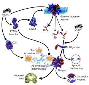
Consult your physician about taking low-dose aspirin or another NSAID concurrently with amphetamine. Non-steroidal anti-inflammatory drug (NSAID) use reduces risk of the development of Parkinson’s disease, protects against 6-hydroxydopamine (6-OHDA) neurotoxicity and Adderall neurotoxicity to dopaminergic neurons in animal models5.
Possible mechanisms include NSAID inhibition of the enzyme cyclooxygenase in the brain which synthesizes protraglandins among other inflammatory mediators; the neurotoxin 6-OHDA elicits large elevations in prostaglandins. Moreover, acetominophen and aspirin inhibit superoxide anion generation and lipid peroxidation, and protect against 1-methyl-4-phenyl pyridinium (MPTP)-induced dopaminergic neurotoxicity in rats6.
N-methyl-D-Aspartate Receptor (NMDAR) Antagonists
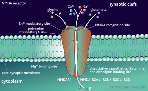
Supplement with magnesium religiously. It has been appreciated for decades that many N-Methyl-D-Aspartate (NMDA) receptor antagonists protect against 6-hydroxydopamine, methamphetamine and adderall neurotoxicity. NMDA receptors bind glutamate and play an important role in learning and memory.
NMDA antagonists include MK-801, dextromethorphan hydrobromide (DXM HBR) and the Alzheimer’s drug memantine (Nameda) [7].
Under physiologic conditions, the divalent cation magnesium occludes the pore of the NMDAR, preventing calcium influx unless a depolarization event pushes the ion away from the channel. Hence, magnesium behaves like a weak endogenous NMDAR antagonist, and has been administered intravenous in emergent situation to help protect the brain against injury. Memantine, another NMDAR antagonist, has also been used successfully to reverse stimulant tolerance. The precise link between NMDAR-dependent excitotoxicity and Adderall neurotoxicity requires further clarification.
Melatonin
Supplement with extended-release melatonin available from PowderCity in the evening. Melatonin is an endogenous neurohormone and chronobiotic produced by the pineal gland which plays important roles in sleep/wake regulation and circadian rhythm.
Melatonin is also among the most potent antioxidants, and has been reported to inhibit 6-OHDA production in vivo, protect against experimental parkinsonism in rodents and attenuate METH and Adderall neurotoxicity [8]. Since melatonin has a short half-life and is rapidly cleared from the CNS, greater benefit will be obtained with extended-release formulations of melatonin.
Pretreatment with lower psychostimulant doses is neuroprotective
If you are psychostimulant-naive and are thinking about initiating psychostimulant therapy to control inattentive symptoms of ADHD, start at the lowest possible dose and titrate upward slowly. Recent evidence has emerged that pretreatment with low-dose AMP and METH in young animals ameliorates the neurotoxicity of future high dose challenge with these drugs [9]. Lower doses may give the brain time develop an adequate defense against future insults.
Avoid concurrent alcohol use
Abstain from alcohol use when taking psychostimulants. Dopamine catabolism is initiated by oxidative deamination via monoamine oxidase to yield 3,4-dihydroxyphenylacetaldehyde (DOPAL). DOPAL is widely-known to be a toxic and reactive intermediate. DOPAL is metabolized primarily via aldehyde dehydrogenase enzymes to a less toxic acid species.
According to Doorn et al 10, aldehyde dehydrogenase inhibition promotes the formation of this reactive dopamine metabolite which is neurotoxic to dopaminergic neurons. Ethanol or alcohol is also a substrate for aldehyde dehydrogenase, which oxidizes ethanol to acetaldehyde, the toxic compound that causes the symptoms of a hangover. Therefore, concurrent alcohol use would theoretically potentiate both METH and Adderall neurotoxicity by competitively inhibiting aldehyde dehydrogenase enzymatic activity, leading to an accumulation of the reactive and autotoxic intermediate 3,4-dihydroxphenylacetaldehyde (DOPAL).
N-Acetyl-Cysteine (NAC) and Selenium
Supplement with NAC and selenium. METH and AMP both deplete endogenous antioxidant defense and intracellular glutathione, and oxidative stress plays an important role in the neurotoxicity of these substances. N-Acetyl-Cysteine serves as a prodrug to the amino acid L-cysteine, which is a precursor to the antioxidant glutathione, and hence administration with N-Acetyl-Cysteine replenishes glutathione stores.
Although selenium is toxic in large doses, in small doses it is an essential micronutrient and functions as a cofactor for reduction of antioxidant enzymes such as glutathione peroxidase, a family of enzymes that catalyze reactions to remove reactive oxygen species. It is likely that NAC and selenium supplementation ameliorates METH and Adderall neurotoxicity by bolstering antioxidant defense and replenishing glutathione stores 11.
Green tea phytochemicals
Swallow your daily dose of adderall with a cup of hot green or white tea. Phytochemicals are naturally abundant compounds that occur in plants, including polyphenols and flavonoids. These are potent agents that mitigate neurodegenerative disease by scavenging pathological concentrations of reactive oxygen species (ROS), reactive nitrogen species (RNS), in addition to chelating neurotoxic transition metals.
Hence, it is not so surprising that phytochemicals protect glia and dopaminergic neurons from adderall neurotoxicity, in addition to the neurotoxins MPTP, rotenone and 6-OHDA.
Specifically, the flavonol (-)-epigallocatechin-3-gallate (EGCG), flavone baicalein, and isothiocyanate sulforaphane (SFN) have demonstrated robust neuroprotective effects [12]. Ruan H et al have also reported that (+/-)-catechin treatment protected against MPTP-induced death of dopaminergic neurons in the substantia nigra and restored striatial dopamine, which is typically depleted after MPTP exposure. The authors speculated that the neuroprotective effects of (+/-) catechin were related to the suppression of c-Jun N-terminal kinase (JNK) and glycogen synthase kinase (GSK) 3beta signaling cascades. Almajano MP’s group also demonstrated a neuroprotective effect of white tea *in vitro *against oxidative stress 13.
Aggressive (and potentially high-risk) Neuroprotective Strategies
10. Minocycline
In animal models, minocycline is probably the single most robust neuroprotective agent identified to-date. Hundreds of peer-reviewed papers have been published elucidating minocycline’s neuroprotective effects. Minocycline has been used for decades as an antibiotic, but it has recently been repurposed in psychiatry and neurology as a treatment for everything from schizophrenia to neurodegenerative disease due to its pleiotropic neuroprotective properties141516.
Minocycline directly inhibits apoptosis (programmed cell death), microglial activation, and a wide variety of inflammatory mediators, including tumor necrosis factor alpha (TNFa), NF-kappa-B and other cytokines. Moreover, minocycline inhibits inducible nitric oxide synthase (NOS). Nitric oxide signaling plays a well-defined role in Adderall neurotoxicity17. Minocycline also modulates glutamatergic signaling in a manner that protects against excitotoxicity. Minocycline use is not risk-free, however. Rare adverse effects include turning the skin blue, hepatic impairment (elevated transaminases), drug-induced lupus and intracranial hypertension due to perturbations in brain osmotic balance.
References
-
Curtin K, Fleckenstein AE, Robison RJ, Crookston MJ, Smith KR, Hanson GR. Methamphetamine/amphetamine abuse and risk of Parkinson’s disease in Utah: a population-based assessment. Drug Alcohol Depend. 2015;146:30-8. ↩
-
Angelucci F, Gruber SH, El khoury A, Tonali PA, Mathé AA. Chronic amphetamine treatment reduces NGF and BDNF in the rat brain. Eur Neuropsychopharmacol. 2007;17(12):756-62. ↩
-
Spencer TJ, Brown A, Seidman LJ, et al. Effect of psychostimulants on brain structure and function in ADHD: a qualitative literature review of magnetic resonance imaging-based neuroimaging studies. J Clin Psychiatry. 2013;74(9):902-17. ↩
-
Wang JY, Wu JN, Cherng TL, et al. Vitamin D(3) attenuates 6-hydroxydopamine-induced neurotoxicity in rats. Brain Res. 2001;904(1):67-75 ↩
-
Carrasco E, Casper D, Werner P. Dopaminergic neurotoxicity by 6-OHDA and MPP+: differential requirement for neuronal cyclooxygenase activity. J Neurosci Res. 2005;81(1):121-31. ↩
-
Maharaj DS, Saravanan KS, Maharaj H, Mohanakumar KP, Daya S. Acetaminophen and aspirin inhibit superoxide anion generation and lipid peroxidation, and protect against 1-methyl-4-phenyl pyridinium-induced dopaminergic neurotoxicity in rats. Neurochem Int. 2004;44(5):355-60. ↩
-
Ohmori T, Koyama T, Muraki A, Yamashita I. Competitive and noncompetitive N-methyl-D-aspartate antagonists protect dopaminergic and serotonergic neurotoxicity produced by methamphetamine in various brain regions. J Neural Transm Gen Sect. 1993;92(2-3):97-106. ↩
-
Borah A, Mohanakumar KP. Melatonin inhibits 6-hydroxydopamine production in the brain to protect against experimental parkinsonism in rodents. J Pineal Res. 2009;47(4):293-300. ↩
-
Hodges AB, Ladenheim B, Mccoy MT, et al. Long-term protective effects of methamphetamine preconditioning against single-day methamphetamine toxic challenges. Curr Neuropharmacol. 2011;9(1):35-9. ↩
-
Doorn JA, Florang VR, Schamp JH, Vanle BC. Aldehyde dehydrogenase inhibition generates a reactive dopamine metabolite autotoxic to dopamine neurons. Parkinsonism Relat Disord. 2014;20 Suppl 1:S73-5. ↩
-
Zhang X, Banerjee A, Banks WA, Ercal N. N-Acetylcysteine amide protects against methamphetamine-induced oxidative stress and neurotoxicity in immortalized human brain endothelial cells. Brain Res. 2009;1275:87-95. ↩
-
Kita T, Asanuma M, Miyazaki I, Takeshima M. Protective effects of phytochemical antioxidants against neurotoxin-induced degeneration of dopaminergic neurons. J Pharmacol Sci. 2014;124(3):313-9. ↩
-
Almajano MP, Vila I, Gines S. Neuroprotective effects of white tea against oxidative stress-induced toxicity in striatal cells. Neurotox Res. 2011;20(4):372-8. ↩
-
Tikka T, Fiebich BL, Goldsteins G, Keinanen R, Koistinaho J. Minocycline, a tetracycline derivative, is neuroprotective against excitotoxicity by inhibiting activation and proliferation of microglia. J Neurosci. 2001;21(8):2580-8. ↩
-
Plane JM, Shen Y, Pleasure DE, Deng W. Prospects for minocycline neuroprotection. Arch Neurol. 2010;67(12):1442-8. ↩
-
Elewa HF, Hilali H, Hess DC, Machado LS, Fagan SC. Minocycline for short-term neuroprotection. Pharmacotherapy. 2006;26(4):515-21. ↩
-
Di monte DA, Royland JE, Jakowec MW, Langston JW. Role of nitric oxide in methamphetamine neurotoxicity: protection by 7-nitroindazole, an inhibitor of neuronal nitric oxide synthase. J Neurochem. 1996;67(6):2443-50. ↩
-
Miyazaki I, Asanuma M. Dopaminergic neuron-specific oxidative stress caused by dopamine itself. Acta Med Okayama. 2008;62(3):141-50. ↩
-
State-based Prevalence Data of Parent Reported ADHD Diagnosis by a Health Care Provider ↩


